
11 Jan Musculoskeletal Ultrasound and Enthesophytes
Neuromuscular ultrasound is a diagnostic imaging technique that uses sound waves to produce images of muscles, tendons, nerves, and other soft tissues. It can be used to help diagnose neuromuscular disorders, such as muscle tears, nerve compressions, and muscle atrophy.
Enthesophytes, also known as bone spurs, are bony growths that can occur at the attachment sites of tendons and ligaments to the bones. They typically develop as a result of chronic stress or inflammation at these attachment sites.
Diagnosing enthesophytes can be done using various imaging techniques, including X-rays, ultrasound, and MRI. Ultrasound is particularly useful for assessing the tendons and ligaments at their insertion points, where enthesophytes commonly occur. The ultrasound can help visualize the bony growths and can also evaluate the surrounding soft tissues for signs of inflammation or other abnormalities.
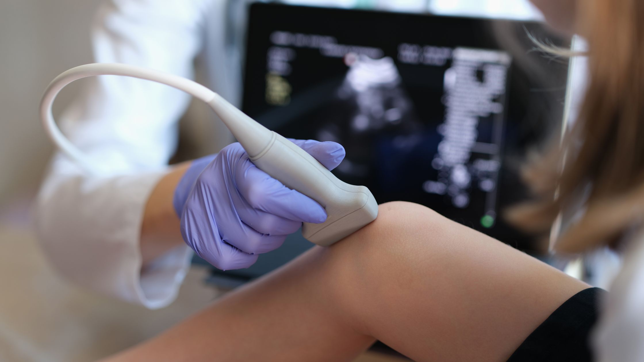
During the ultrasound examination, the physician or technician will apply a gel to the skin and use a handheld device called a transducer to emit sound waves into the body. The sound waves then bounce back and are converted into images that can be seen on a monitor. By moving the transducer along the area of interest, the healthcare professional can obtain detailed
images of the tendon and its attachment site, allowing for identification of any enthesophytes present.
The diagnosis of enthesophytes is based on the findings seen on the ultrasound images. Th.ese bony growths appear as hyperechoic, or bright, structures attached to the bone at the tendon insertion site. Depending on their size and shape, they may be classified as either small, smooth enthesophytes or larger, irregular enthesophytes. In addition to the presence of the enthesophytes, the ultrasound may also show signs of inflammation, such as thickening or hypoechoic, or dark, areas around the tendon insertion.
In summary, neuromuscular ultrasound can be used to diagnose enthesophytes by visualizing the bony growths at the tendon insertion site. This imaging technique provides a detailed assessment of the tendons, ligaments, and surrounding soft tissues, allowing healthcare professionals to identify and evaluate the presence of enthesophytes and any associated inflammation